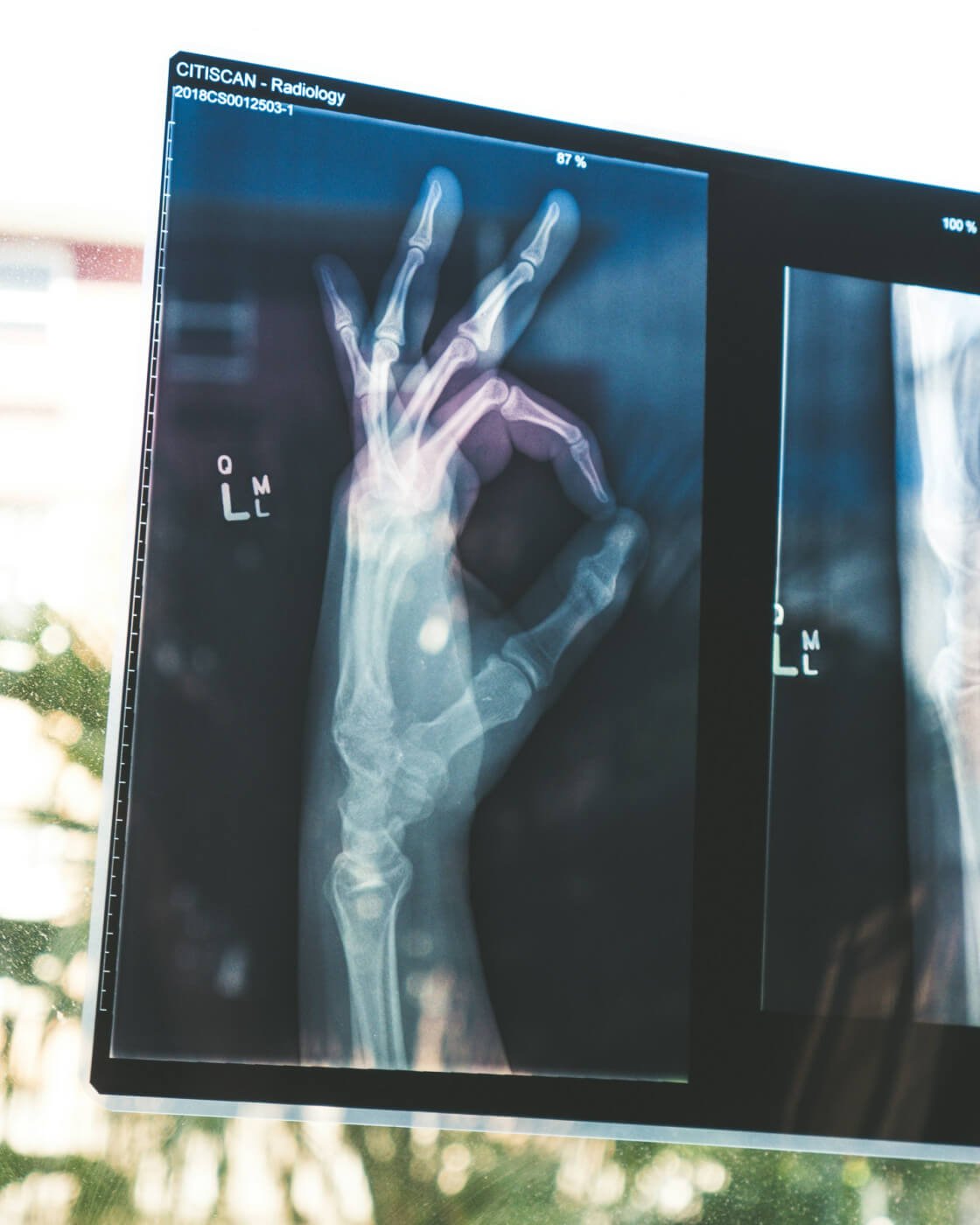
An X-Ray Marker is made of lead and shows up on X-Rays to indicate the side of the body being imaged. In English L and R stand for Left and Right respectively.
Often institutions will ask their Radiographers to include their initials on the X-Ray markers, or a number that correlates to their name within the Radiology department. This ensures the Radiographer who took the X-Ray remains accountable for their work, and if any follow up is required the person performing the examination can easily be found.
Students are sometimes required to provide their initials and an indicaiton of the University they attend on their markers that are used on clinical placments. Some Universities only require a Left and Right marker and initials are not required until the Radiographer graduates. It all depends on where you work and what the expectation is within that particular department.
It is important to correctly display which side of the patient is imaged to the radiologist and the referring team. If the patient requires follow up such as surgery for example; if the incorrect side is labelled on the X-Ray, it is possible that may lead to mistakes identifying where the fracture is further down the patient care pathway.

We have the largest range of options worldwide for you to customise your own X-Ray Markers here.
Custom X-Ray Markers

$20.00
Create your own unique set of X-Ray Markers, add your initials if required, choose your shape, size and colour from our extensive range of 250 glitters! One set of left and right x-ray markers made the way you like! Want… read more
You can include your own initials, choose colours and pick your preferred size.
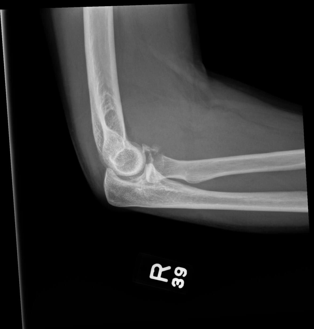
The image displayed is a Right Lateral Elbow X-Ray with a Coronoid Process Fracture. The 39 under the R correlates with the identity of the staff member who took the Radiograph.
Image courtesy of Radiopaedia, an online learning tool for all things Radiology.
Case courtesy of Kevan English, <a href="https://radiopaedia.org/?lang=us">Radiopaedia.org</a>. From the case <a href="https://radiopaedia.org/cases/181127?lang=us">rID: 181127</a>